AHNAK2 Antibody (PACO58272)
- SKU:
- PACO58272
- Product type:
- Antibody
- Reactivity:
- Human
- Host Species:
- Rabbit
- Isotype:
- IgG
- Application:
- ELISA
- Application:
- IHC
- Application:
- IF
- Antibody type:
- Polyclonal
- Conjugation:
- Unconjugated
Frequently bought together:
Description
| Antibody Name: | AHNAK2 Antibody (PACO58272) |
| Antibody SKU: | PACO58272 |
| Size: | 50ug |
| Host Species: | Rabbit |
| Tested Applications: | ELISA, IHC, IF |
| Recommended Dilutions: | ELISA:1:2000-1:10000, IHC:1:200-1:500, IF:1:50-1:200 |
| Species Reactivity: | Human |
| Immunogen: | Recombinant Human Protein AHNAK2 protein (195-349AA) |
| Form: | Liquid |
| Storage Buffer: | Preservative: 0.03% Proclin 300 Constituents: 50% Glycerol, 0.01M PBS, pH 7.4 |
| Purification Method: | >95%, Protein G purified |
| Clonality: | Polyclonal |
| Isotype: | IgG |
| Conjugate: | Non-conjugated |
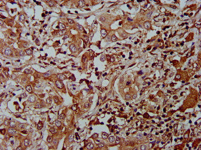
 | IHC image of PACO58272 diluted at 1:400 and staining in paraffin-embedded human liver cancer performed on a Leica BondTM system. After dewaxing and hydration, antigen retrieval was mediated by high pressure in a citrate buffer (pH 6.0). Section was blocked with 10% normal goat serum 30min at RT. Then primary antibody (1% BSA) was incubated at 4°C overnight. The primary is detected by a biotinylated secondary antibody and visualized using an HRP conjugated SP system. |
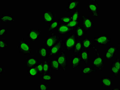
 | Immunofluorescence staining of A549 cells with PACO58272 at 1:133, counter-stained with DAPI. The cells were fixed in 4% formaldehyde, permeabilized using 0.2% Triton X-100 and blocked in 10% normal Goat Serum. The cells were then incubated with the antibody overnight at 4°C. The secondary antibody was Alexa Fluor 488-congugated AffiniPure Goat Anti-Rabbit IgG(H+L). |
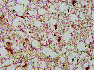
 | IHC image of PACO58272 diluted at 1:400 and staining in paraffin-embedded human brain tissue performed on a Leica BondTM system. After dewaxing and hydration, antigen retrieval was mediated by high pressure in a citrate buffer (pH 6.0). Section was blocked with 10% normal goat serum 30min at RT. Then primary antibody (1% BSA) was incubated at 4°C overnight. The primary is detected by a biotinylated secondary antibody and visualized using an HRP conjugated SP system. |
| Background: | costamere, cytoplasm, cytoplasmic vesicle membrane, cytosol, plasma membrane, sarcolemma, T-tubule, Z disc |
| Synonyms: | Protein AHNAK2, AHNAK2, C14orf78 KIAA2019 |
| UniProt Protein Function: | |
| UniProt Protein Details: | |
| NCBI Summary: | This gene encodes a large nucleoprotein. The encoded protein has a tripartite domain structure with a relatively short N-terminus and a long C-terminus, separated by a large body of repeats. The N-terminal PSD-95/Discs-large/ZO-1 (PDZ)-like domain is thought to function in the formation of stable homodimers. The encoded protein may play a role in calcium signaling by associating with calcium channel proteins. Alternative splicing results in multiple transcript variants. [provided by RefSeq, Apr 2017] |
| UniProt Code: | Q8IVF2 |
| NCBI GenInfo Identifier: | 113424746 |
| NCBI Gene ID: | 113146 |
| NCBI Accession: | XM_290629 |
| UniProt Secondary Accession: | Q8IVF2 |
| UniProt Related Accession: | Q8IVF2 |
| Molecular Weight: | |
| NCBI Full Name: | |
| NCBI Synonym Full Names: | AHNAK nucleoprotein 2 |
| NCBI Official Symbol: | AHNAK2 |
| NCBI Official Synonym Symbols: | C14orf78 |
| NCBI Protein Information: | protein AHNAK2 |
| UniProt Protein Name: | |
| UniProt Synonym Protein Names: | |
| Protein Family: | Protein |
| UniProt Gene Name: | |
| UniProt Entry Name: |


